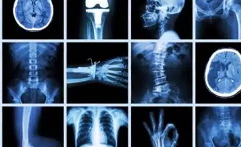
The key to a guided injection is to get a look inside your joint or your spine without any surgery at all. That’s where X-ray imaging with fluoroscopy comes in handy. The special machine used is portable and is shaped like a “C” – giving it the name the C-arm. This machine can be rotated around your body to get the best angle on the joint or spot in your spine that’s the source of your pain.
During your exam, continuous X-ray images will give your doctor real-time information. That means if you’re getting an injection, your doctor is actually watching the needle as it is inserted and knows when the needle is in the right place inside your joint. This live information helps deliver guided therapeutic injections that can relieve your pain.
Once you’re checked in and changed, our technologist will help you get into the right position on the X-ray table. The C-arm will be rolled into place and rotated to the best angle to image your joint or spine. Then your skin will be cleaned around the image area and numbed with a local anesthetic.
For some exams, a contrast material may be injected as a next step. This contrast can help highlight an area and make it possible for your doctor to see internal organs and tissues in motion. To inject the contrast, we’ll use a thin needle. You can expect to feel pressure or brief discomfort. If a therapeutic pain injection is part of your procedure, your doctor will use the X-ray guidance to precisely position the needle.
We’ll move the C-arm out of the way when you’re finished and help you get up from the table. Certain procedures also require more imaging. Depending on your exam, you may be taken to the MRI suite, the CT room, or another exam room for more X-ray.
Finding the source of joint pain joint pain and making a diagnosis is what an Arthrogram is all about. Whether it’s a hip, a shoulder, or any other joint, getting real-time imaging to help guide a thin needle into your joint can help get you answers.
Musculoskeletal Radiologist Keith Baynes explains, “The hard thing about all of imaging is we don’t image pain. Patients come in with a symptom, they come in with pain. We do an X-ray, we do an MRI, and they say, ‘Where’s my pain coming from?’ That’s very difficult because the pain doesn’t light up on the image. You have to find the structure that relates to the patient’s pain.”
Your exam starts with getting you positioned on the X-ray table. You’ll be numbed up and then fluoroscopy is used to guide the needle into your joint where contrast is injected. That contrast distends or blows the joint up from the inside, kind of like a balloon. Most patients feel tightness or fullness in the joint, but not pain. Your next stop is more imaging, which you’ll get right away because your body will absorb the contrast within 20 or 30 minutes. An Arthrogram is the best non-surgical option for examining joints and understanding the underlying cause of your pain.
For more on what it’s like to get an Arthrogram, click here.
For more on getting an Arthrogram with MRI, click here.
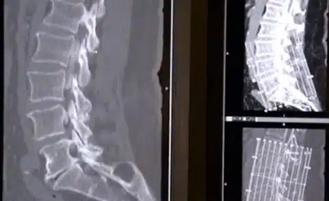
To pinpoint and treat back pain involving discs, your doctor may recommend a Discogram. This procedure is done with fluoroscopy in the C-arm room. The goal is to find the source of your pain. Once you are positioned on the X-ray table and the C-arm is in place, your doctor will use fluoroscopy to carefully place a needle into the center of your disc.
During your exam, you might be asked, “Is this the kind of pain that you’re suffering that brought you to the doctor?” Then, if painful, a strong local anesthetic is injected right into the disc to relieve that pain. Within a minute of the injection, most patients’ pain is relieved.
Depending on your pain, you may need injections in several discs. Patients who have tried other treatments, medications and therapies and have been suffering for months or years, are able to walk out of the appointment feeling immediate relief.
Click here to learn how a discogram can find the source of your chronic back pain.
An exam called a Myelogram uses fluoroscopy and other imaging to help diagnose back pain. For RAYUS Radiology patient Linda Zentgraf-Kierski, her back pain was getting so intense she began worrying she’d spend her life in a wheelchair. “I pick up my grandkids five days a week and I’m not going to be able to pick them up,” she explained before her Myelogram
Linda’s exam started in the C-arm room where her doctor will used fluoroscopy to confirm the needle is in the right spot, then inject contrast into the source of the chronic pain. Linda described the contrast injection as a cold tingle. She was then moved to an X-ray machine for about 10 minutes of imaging. Finally, she went to the CT scanner for approximately 5 more minutes of scans. Linda’s last stop was the recovery room where she rested under the watchful eye of nurses for about two hours.
The goal of the contrast injection and all of the imaging is to give her surgeon a clear picture of what is happening in her spine. With that information, she and her surgeon can make the best plan for treatment, and ultimately for relief so Linda can get back to her active life with her grandkids.